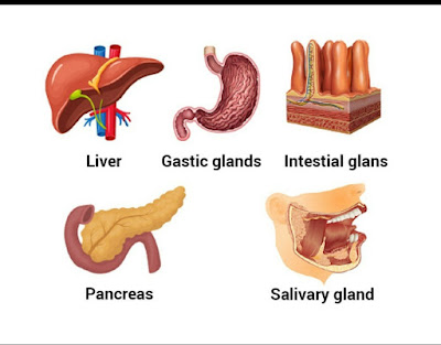
- 1. SALIVARY GLANDS
- 2. GASTRIC GLANDS
- 3.INTESTINAL GLANDS
- 4.LIVER
- 5.PANCREAS
1. SALIVARY GLANDS:- Three pairs of salivary glands are seen in humans. However, in some mammals like rabbits, 4 pairs of salivary glands are seen, i.e., they also include INFRA-ORBITAL GLANDS also. The 3 pairs of salivary glands in human are;
A. PAROTID GLAND:- These are present at below of pinna/inner surface of cheeks.
B. SUB-MAXILARY GLAND:- These gland is also called as sub-mandibular gland. These are present at angles of lower jaw.
C.SUB-LINGUAL GLANDS:- These are present below the tongue. These glands are situated just outside the buccal cavity.
The salivary glands have mucosa and serosa cells. Mucus is secreted by mucosa cells. Salivary amylase is secreted by serosa cells. Saliva have 99.5% of water and 0.5% of solutes (such as ions, IgA antibodies, ptylalin or alpha amylase and lysozyme.) This lysozyme kills bacteria. The ph of saliva is 6.8.
2. GASTRIC GLANDS:- These are located at stomach beneath tje surface epithelium. Here we see 3 types of glands;
A. CARDIAC GLANDS:- These secrete mucus.
B. PYLORIC GLANDS:- Secrete mucus and harmone GASTRIN.
C. FUNDARIC GLANDS:- They are composed of 3 different cells.
- i. NECK CELLS:- They secrete mucus.
- ii. PEPTIC CELLS:- These cells used to secrete large amount of pro-enzymes i.e., pepsinogen, prorenin.
- iii. OXYNTIC CELLS:- These produce hydrochloric acid.
Secretion of these glands from gastric juice ph from 0.9 to 1.8.
3. INTESTINAL GLANDS:- These glands are of two types;
INTESTINAL GLANDS OF HUMAN
A. BRUNNER'S GLANDS:- These are located at sub-mucosa of duodenum and secrete alkaline mucus.
B. CRYPTS OF LIEBERKUHN:- These are located at mucosa of small intestine and secrete intestinal juice/succus entericus.
This intestinal juice have ph 7.8. This juice have carbohydrates like carbohydrate digestive enzymes called maltase, sucrase (invertase) and lactase. This juice also have protein digestive enzymes called peptidases like amino-peptidases, tripeptidases, and dipeptidases, nucleo-peptidases and small amount of intestinal lipase and enzyme activator enterokinase. The wall of intestine have STEM CELLS which play role in renewing the epithelial cells lost in intestinal epithelium. Paneth cells believed to play a role in protection of those cells.
4. LIVER:- It is the largest gland of our body. It's weight is about 1.2 to 1.5 kg in an adult human. It is situated at right side of abdominal cavity, just below the diaphragm.
*ANATOMY:- It is a reddish brown organ of 4 unequal lobes. The two principal lobes of liver are divided bu a fold of mesentary.
*HISTORY:- Each lobe is divided into many hexagonal lobules. These hepatic lobules are structural and functional units of liver. Each lobule is covered by the connective tissue sheath called as GLISSON'S CAPSULE. The hepatocytes in liver lobule form a series of irregular cords called as HEPATIC TREBECULAE, it is arranged like spokes of a wheel. The lining of sinusoids have large no.of KUPFFER CELLS, which are also called as stellate reticulo endothelial cells. A typical liver lobule is hexagonal in cross section and these are 6 portal triads one at each corner of the lobule. A portal triad have branch of hepatic portal vein, a branch of hepatic artery, small branch of bile duct. Hepatocytes secrete BILE into BILE CANALICULI and they merge to form BILE DUCTS. These bile ducts eventually merge to form left and right hepatic ducts which unite and exit liver as common HEPATIC DUCT. The bile passes through hepatic ducts and it is stored and concentrated in a thin muscular sac called GALL BLADDER. The duct of gall bladder along with hepatic duct from liver forms the common bile duct.
*BILE:- The bile has ph 7.6 to 8.6. This contains water, bile salts, lecithin, bile pigments, cholesterol, etc; The bile has no enzymes. The bile salts help in EMULSIFICATION of fats.
FUNCTIONS OF LIVER:- Liver performs various of functions like storage, secretion and synthesis of various substances. Carbohydrate metabolism, detoxification, lipid metabolism, protein metabolism,thermogenesis,haemopoiesis,erythroclasia, phagocytosis,
synthesis of clotting factors and etc:-
5. PANCREAS:- It is a pinkish grey colored elongated organ. Second largest gland in the body. It is posterior to stomach within curvature of duodenum. It have a head, body and tail. Head lies with in duodenal loop. Slender body extends towards spleen. Tail is short and bluntly rounded. Pancreas is both exocrinic and endocrinic organ
and the exocrinic portion secretes an alkaline pancreatic juice which contains enzymes. The large pancreatic duct and common bile duct join to form HEPATO-PANCREATIC AMPULLA or AMPULLA OF VATER. The opening of ampulla into duodenum on major duodenal papilla or regulated by SPHINCTER OD ODDI. ISLETS OF LANGERHANS are present as endocrinic tissues of pancreas. These are secreted as ALPHA and BETA CELLS. These are scattered among ACINI. Alpha cells secrete GLUCAGON and beta cells secrete INSULIN into blood stream to regulate blood glucose levels.
HUMAN PANCREAS
These are the 5 major glands useful for the digestion.


Comments
Post a Comment